PorCORA
Porcine Cornea Opacity/Reversibility Assay: An Alternative to the Draize Ocular Irritation Assay

Introduction
MB Research has developed the Porcine Cornea Opacity/Reversibility Assay (PorCORA) – an alternative testing protocol for reversibility of ocular damage in excised porcine corneas. The ex vivo corneal epithelium is dosed topically with a test substance, similar to the BCOP Test, but by culturing the excised corneas for up to 21 days, the potential for the damage reversal can be assessed.
The Missing Puzzle Piece
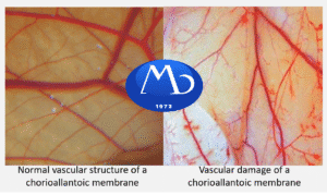
There are several alternative methods to characterize aspects of eye irritation and damage, but no established method can model recovery after injury like the traditional Draize test. The PorCORA was developed to fill this void by measuring corneal damage and recovery for extended periods in excised porcine corneas. Combined with other alternative assays, such as the HET-CAM or CAMVA for assessment of conjunctival injury and vascular damage and the BCOP Test for assessment of acute ocular irritation, the PorCORA is the missing piece that allows for evaluation of corneal healing.
The PorCORA evaluates similar mechanisms to those occurring in an in vivo Draize assay, in which test substances are placed directly onto corneal tissue. Any damage to the tissue is visualized by the retention of fluorescein. Tissues can be repeatedly stained with fluorescein to assess changes in the area of damage over time; therefore, the potential of tissue recovery can be monitored. Because corneal damage is generally the most persistent of severe ocular injuries, the time course measurements in the PorCORA are justifiable in modeling both reversible and non-reversible eye damage.
Current Validation Developments
Tracking Reversibility
MB Research Scientists have developed assay methods in order to create a cost-effective protocol as a viable alternative to the Draize Rabbit Eye Test. In validation experiments, excised corneas have been monitored for over three weeks with minimal morphological changes to the corneal epithelium.
Re-epithelialization of the corneal surface (reversibility) can be measured with fluorescein stain, which is representative of actual tissue damage determined by fluorescence confocal microscopy (live/dead stain), reflective confocal microscopy (no stain), and histology.
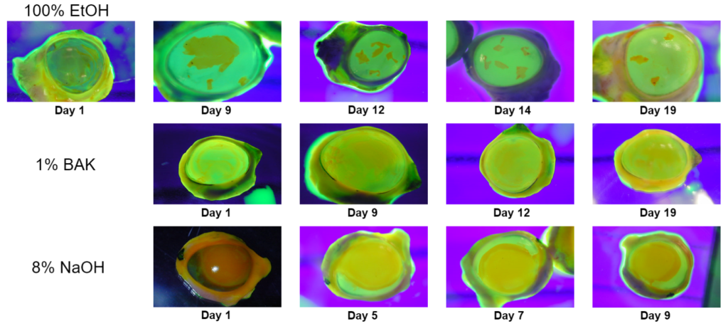
The photos below show the area of fluorescein stain retention of a cornea treated with 100% ethanol (EtOH) decreasing over time indicating reversibility. In 8% Sodium Hydroxide (NaOH)-treated corneas, the area that retained the stain did not decrease over time, indicating irreversible damage. Although the corneas treated with 1% Benzalkonium chloride (BAK) showed some recovery, significant damage was still observed at Day 19. No significant fluorescein retention was visualized in corneas treated with Dulbecco’s phosphate-buffered saline (DPBS) (images not shown). These results show PorCORA’s potential to discriminate eye irritants that cause reversible damage (100% EtOH) from those that irreversibly damage corneal epithelium (8% NaOH).

Confocal Microscopy and Live/Dead Staining.
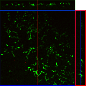
To confirm recovery of corneal epithelial cells post-dose administration in the PorCORA, cell viability was visualized by using confocal microscopy in conjunction with a live/dead fluorescent staining kit.
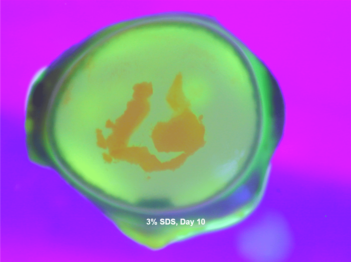
Corneal cell viability was consistent with typical rates of recovery from Sodium dodecyl sulfate (SDS) treatment seen by fluorescein retention. A dense area of live cells was observed localized adjacent to a dense area of dead cells on a recovering corneal surface.
Confocal microscopy results support the current model of corneal recovery by epithelial sheet migration. On Day 10, the SDS-treated corneas were populated with many live epithelial cells and shows that re-epithelialization occurs during the time course of this culture. Similar validations were performed in DPBS.
3% SDS Day 3
Optical sectioning through the corneal surface shows extensive cell death and few live cells.


3% SDS Day 10
Day 10 SDS-treated corneas are populated by numerous live cells, and very few dead cells, demonstrating reversibility and recovery of tissue damage in this corneal culture model.
PorCORA Predicts EU R41 and GHS Category 1
Since some regulatory classification methods of ocular irritancy depend upon the time for an ocular injury to completely heal (OECD, 1967; WHMIS, 1988; HMIS, 1996; EPA, 1997), PorCORA was developed to measures ocular irritation and reversibility following chemical insult in excised porcine corneal tissues for extended periods.
Paired with other assays that will quantify the severity (such as the BCOP, Epi-Ocular, and Chorioallantoic membrane-based assays), PorCORA is an excellent method to replace in vivo ocular assays.
This model system is representative of corneal healing in the rabbit eye, as comparison of average days to clear in PorCORA to average days to clear in the ECETOC historical Draize database yields a correlation coefficient of 0.84.
In a validation project consisting of 32-reference compounds, PorCORA yielded very favorable results correctly predicting reversibility (88%), EU: R41 (91%), GHS: Category 1 (91%), and EPA: Category I (88%).


